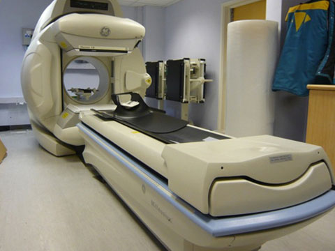
دوربین گاما
دوربین گاما ( Gamma Camera )امروزه یکی از ابزارهای مهم در پزشکی هسته ای دوربین گاما است. این وسیله برای به تصویر کشیدن پرتوهای گامای ساطع شده از عضو هدف بکار می رود. در بخش پزشکی هسته ای به بیمار رادیو ایزوتوپ داده می شود که باعث می شود رادیواکتیویته در بافت هدف تجمع یابد.
این کار از سه طریق تزریق به درون رگ، استنشاقی و یا به صورت خوراکی قابل انجام است. رادیو ایزوتوپ دارای این ویژگی است که در ناحیه ای با متابولیسم بیشتر، تجمع بیشتری پیدا می کند.
دوربین گاما
بعضی از رادیوایزوتوپ ها دارای بافت هدف چندگانه هستند و در اسکن از کل بدن مورد استفاده قرار می گیرند.به عنوان نمونه،TC-Dtap رادیوایزوتوپی است که در ناحیه ی کلیه تجمع پیدا می کند و محل تجمع TC-Dmsa در ناحیه ی کبد است.
درون بافت،رادیو ایزوتوپ شروع به پرتودهی می کند و خود بافت منبع تابش پرتو می شود. به این ترتیب اشعه ی گاما با انرژی مناسببه سمت آشکارساز ساطع می شود.
ساختار دستگاه
دوربین گاما از یک یا چندین صفحه ی کریستال سنتیلاتور تشکیل یافته است.فوتون های آزاد شده از بافت ابتدا از کولیماتور عبور می کنند. که از صفحه ی ضخیمی از جنس سرب به ضخامت حدود 1 الی 3 اینچ تشکیل شده و دارای سوراخ های زیادی است که سنتیلاتور نامیده می شوند. زمانی که تابش یونیزه کننده از درون سنتیلاتور عبور می کند، فوتون هایی را به وجود می آورد که در فوتومولتی پلایر (Photomultiplier) ، که بخشی دارای لایه ای با خاصیت فوتوالکتریک می باشد به سیگنال الکتریکی تبدیل می شوند.
نحوه کار دوربین گاما
الکترون ها در اثر برخورد با فوتوکاتد که با کمک آن ها میدان الکتریکی ایجاد گشته است، تعداد بیشتری الکترون آزاد می نمایند و این روند چندین بار تکرار می گردد تا سطح انرژی الکترون ها به حد مطلوبی برسد. در نهایت پتانسیل های اندازه گیری شده ، توسط تقویت کننده هایی تقویت و ثبت می شوند.
Gamma camera
The gamma camera is the equipment used to detect the distribution of radiopharmaceutical within the patient
Components:
- Collimator
- Radiation detector
- Scintillation crystal
- Photomultiplier tubes
- Electronics
- Preamplifier
Collimator

When radiation is released from the patient it can exit at any angle and hit the detector in a location that doesn’t correlate with the location of its origin. To overcome this a collimator is used in which only gamma photons that travel perpendicular to collimator will be accepted. Those traveling at an angle will hit the septum (usually lead), be absorbed and, therefore, not contribute to the image.
N.B. The collimator acts as a lens to reject photons that have a path that means they do not hit the camera in a location that corresponds to their original location i.e. its purpose is for spatial mapping. It does not reject scatter.
Features of the collimator
Hole direction

- Parallel hole – these are the most common.
- Diverging hole – for a minified image
- Converging hole – for magnifying the image
- Pinhole – single-hole collimator for magnifying images of small objects e.g. thyroid
Hole formation
The holes can be created by:
- Crimped lead foil sheets (cheap but the gaps in the septae degrade image contrast)
- Drilling into a lead block (these give better image contrast as there are no gaps in the septae, but are more expensive)
- Casting from molten lead.
Septal thickness
The higher the energy of the emitted gamma photons the thicker the septae need to be to ensure maximum absorption of photons that hit them at an angle and, therefore, better rejection of non-perpendicular photons. Parallel hole collimators are classified as low, medium or high energy according to their septal thickness.
| Classification | Photon energy (keV) | Septal thickness (mm) | Radionuclide |
| Low energy | 150 | 0.3 | 99mTc |
| Medium energy | 300 | 1 | Indium-111 |
| High energy | 400 | 2 | 131I |

Scintillation crystal

- The crystal is fluorescent i.e. when a gamma photon interacts it releases light photons (mixture of visible and UV light)
- Single crystal of sodium iodide with a small amount of thallium (NaI(Tl)). The thallium improves the light output.
- 6-13 mm thick
- Hermetically sealed in aluminium can
Scatter rejection
If a gamma photon scatters within the patient’s body (via Compton scatter) it will change direction and, therefore, will not hit the detector at a location corresponding to its location of origin. It is important to reject these scattered photons as they degrade the image contrast and spatial resolution. This cannot be done by the collimator and is, therefore, done electronically by a process called energy discrimination.
A gamma photon that scatters within the patient will never hit the scintillator with the full energy (i.e. it won’t lie within the peak). Therefore, only gamma photons in the peak can be confidently identified as non-scattered radiation from the patient.
Usually a 20% acceptance window is used centred on the photopeak. The acceptance window can be adjusted and more than one window can be used for radionuclides that have more than one photopeak (e.g. indium-111 has peaks at 172 and 247 keV). This is made possible by the Z values being displayed with a multi-channel analyser that allows more than one window to be set.
Image formation
Each PMT corresponds to a coordinate on the scintillation crystal. This is then mapped out onto a matrix. Each time a gamma photon that falls within the acceptable energy window is detected it is mapped on to its corresponding coordinate within the image.
Image acquisition is controlled by the user and may be terminated when:
- Preset number of counts obtained
- Preset time passed
Image display
The digital image is displayed upon a monitor with each pixel corresponding to a memory location in the matrix and the brightness / colour scale corresponding to the count number in that location.
Display can be manipulated and optimised by:
- Smoothing to reduce noise
- Windowing to increase contrast
- Interpolation increases the display matrix relative to the acquisition matrix which spreads the counts and makes the pixels less apparent
- Adding and subtracting images to extract quantified information
Pre-amplifier
This converts the current produced at the anode of the PMT to a voltage pulse. The amplitude of the voltage pulse is directly proportional to the charge produced at the anode and, therefore, the amount of light received by the PMT, which is proportional to the number of gamma photons that hit the scintillation crystal.