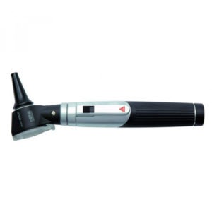
اتوسکوپ(Otoscope)
نام وسیله ای پزشکی است که برای مشاهده ی داخل گوش مورد استفاده قرار می گیرد. پزشکان این وسیله را به منظور غربالگری برخی بیماری ها در معاینات دوره ای معمول و یا بررسی برخی شکایات مرتبط با سیستم شنوایی بکار می برند. با کمک اتوسکوپ می توان گوش خارجی و میانی را مورد معاینه قرارداد.
اتوسکودپ شامل دو بخش «سر» و «دسته» است .«سر» از یک منبع الکتریکی تولید نور و یک عدسی با قدرت بزرگ نمایی کم تشکیل شده است.در انتهای قدامی این وسیله نیز ناحیه ای به منظور اتصال یک اسپکولوم پلاستیکی برای ورود به داخل مجرای گوش در نظر گرفته شده است .
موارد کاربرد اتوسکوپ
وقتی بیماری از درد گوش شکایت دارد برای تشخیص اینکه علت درد،عفونت گوش است و یا از ناراحتی های اعضای مجاور است ، معاینه گوش الزامی است ، بدلیل اینکه مجرای گوش تنها پنجره ای است که به گوش میانی باز می شود و خصوصیات و ظاهروعملکرد پرده ی صماخ اطلاعات مهم و با ارزشی در مورد بیماری های احتمالی گوش میانی در اختیار می گذارد.
انواع اتوسکوپ
دریچه عدسی به صورت متحرک و قابل جداسازی بوده که به وسیله آن می توان برخی از ابزار معاینه را با هدایت اتوسکوپ به مجرای گوش وارد نموند. از این خاصیت به عنوان مثال، در خارج کردن ترشحات گوش (سرومن) استفاده می شود.برخی دیگر از این مدل ها، دارای محلی به منظور وارد کردن هوا از طریق اسپکولوم به داخل مجرا برای ارزیابی میزان تحرک پرده تمپان می باشند.
بسیاری از اتوسکوپ های مورد استفاده در مطب ها،از نوع ثابت بوده، در حالی که برخی نیز قابل حمل هستند. انواع ثابت با کمک یک رابط به منبع الکتریسیته متصل می شوند، در شرایطی که انواع قابل حمل، اغلب حاوی باتری های قابل شارژ هستند.
اتوسکوپ ها می توانند بدون نیاز به کارگذاری اسپکولوم، جداگانه برای معاینه بینی نیز مورد استفاده قرار گیرند .
Learn more about Otoscope
Foreign Body, Ear
Philip Buttaravoli MD FACEP, Stephen M. Leffler MD FACEP, in Minor Emergencies (Third Edition), 2012
What To Do
-
Use an otoscope to inspect the ear canal while pulling up and back on the pinna to help straighten the ear canal, thereby providing a better view.
-
If there is a live insect in the patient’s ear, begin by filling the canal with a liquid to kill the
insect. Mineral oil, 2% lidocaine (Xylocaine), or benzocaine/antipyrine (Auralgan) works well.
(Sterile 2% lidocaine would be most appropriate if there is a myringotomy tube in place or any other opening of the tympanic membrane [TM].)
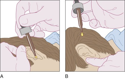
Instruct the patient to lie on his or her side, and then drip the liquid into the canal while pulling
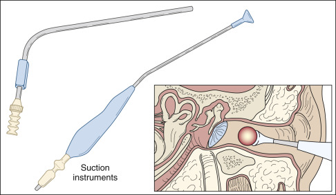
on the pinna and pushing on the tragus to remove air bubbles (Figure 28-1).
Otoscope 0
With a foreign body (FB) that is not too tightly wedged in the canal, and if tympanic membrane
perforation or a myringotomy tube is not present, water irrigation is a very effective removal
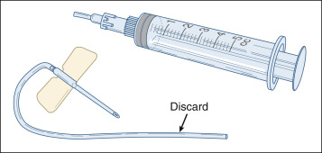
technique. This can be accomplished with a syringe and scalp vein needle that has been cut short (Figure 28-2).
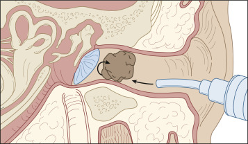
Tap water at body temperature can be used to flush out the foreign body. Direct the stream
along the wall of the canal and around the object, thereby flushing it out (Figure 28-3).
اتوسکوپ 02
Figure 28-2. A short, soft catheter on a 5-mL syringe is safe and very effective for irrigating a loose foreign body out of the ear canal. Note: Discard the tubing after use.
اتوسکوپ 03
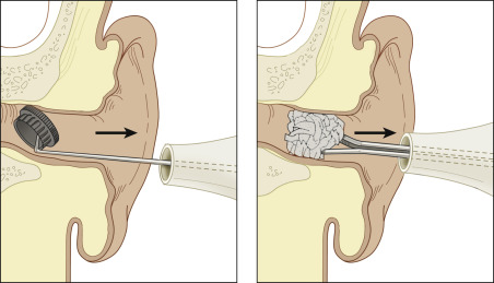
If a hard or spherical object remains tightly wedged in the canal, attempt to roll the foreign body out
by getting behind it with a right-angle nerve hook, ear curette, or wire loop. (Alternatively, a
Calgiswab can be bent into a right-angle hook and used in the same way.) Use of these tools should be done under direct vision through an ear speculum.
اتوسکوپ 04
The patient’s head should be firmly stabilized to prevent sudden movements. Whenever an
instrument is used in the ear canal, warn the patient or parents beforehand that there may be a
small amount of bleeding and pain because of the delicate lining of the ear canal .
An alternative removal technique is to take a drop or two of cyanoacrylate (Super Glue,
Dermabond), and place it on the end of the wooden shaft of a cotton swab. (Use the cotton end for
irregular FBs.) Then hold the wet glue against the foreign object until it hardens (approximately 30
seconds to a minute), and extract the foreign body from the canal.
If the object is light and moves easily, you can attempt to suction it out with a standard metal
suction tip or (if available) a specialized flexible tip, by making an effective vacuum seal on the foreign body .
اتوسکوپ 05
A small magnet or iron-containing metallic foreign body can be removed by touching a pacemaker
magnet to a metal forceps and then, at the same time, touching the forceps to the foreign body,
withdrawing all of the magnetized objects together.
Alligator forceps are good for grasping soft objects, such as cotton, paper, and certain insects.
There should be no delay in removing a foreign body in a canal when there is an obvious
infection or when the foreign body is a disk or button battery. Do not irrigate or instill liquids
into the ear canal, because on contact with moist tissue, the alkaline battery is capable of producing
a liquefactive necrosis extending into deep tissues within hours. Be careful not to crush the battery.
After removal of a battery, irrigate the canal to remove any alkali residue.
At any time, if a child becomes uncooperative, especially when using metal instruments,
use procedural sedation as described in Appendix E. Ketamine sedation appears to have a
positive effect on the success rate of foreign body removal in children.
If the foreign body is tightly wedged in the canal and you cannot remove it, consult an
ear-nose-throat (ENT) specialist. If after removal, there is evidence of infection or perforation of
the tympanic membrane, referral to an ENT specialist is also appropriate.