سونوتراپی
سونوتراپی عبارت است از درمان درد ها و سایر مشکلات با استفاده از امواج اولتراسوند. استفاده از امواج اولتراسوند در درمان پیش از
استفاده ی آن در تشخیص مطرح بوده است. همانطور که می دانید امواج اولتراسوند امواج مکانیکی می باشند و لذا این ارتعاشات الکتریکی
درون بافت باعث تولید حرارت می شوند و این حرارت است که می تواند تسکین دهنده باشد. با استفاده از روش سونوتراپی ما می توانیم بافت
های نیمه عمقی نظیر مفاصل، تاندون ها، لیگامان ها، عضلات و … را درمان کنیم.
همچنین این روش در ترمیم شکستگی استخوان نیز کاربرد زیادی دارد. از مواردی که سونوتراپی به صورت درمان اختصاصی به کار می رود
می توان به درمان فلج عضلات صورت اشاره کرد. در این بیماری به دلیل اینکه استفاده از روش های معمول فیزیوتراپی باعث تبخیر آب چشم
می شوند می توان به راحتی با استفاده از سونوتراپی حرارت را فقط در عمق مورد نظر ایجاد نمود.
سونوتراپی
کاربرد گرمایی سونوتراپی
با جذب امواج فراصوت به وسیله ی بدن بخشی از انرژی آن به گرما تبدیل می شود .گرمای موضعی حاصل از جذب امواج فراصوت بهبودی را
تسریع می کند. قابلیت کشسانی کلاژن ( پروتئینی ارتجاعی ) را افزایش می دهد. کشش در scars ( اسکار= جوشگاه های زخم ) افزایش می
دهد و باعث بهبود آن ها می شود. اگر اسکار به بافت های زیرین خود چسبیده باشد، باعث آزاد شدن آن ها می شود. گرمای حاصل از امواج
فراصوت با گرمای حاصل از گرمایش متفاوت است .
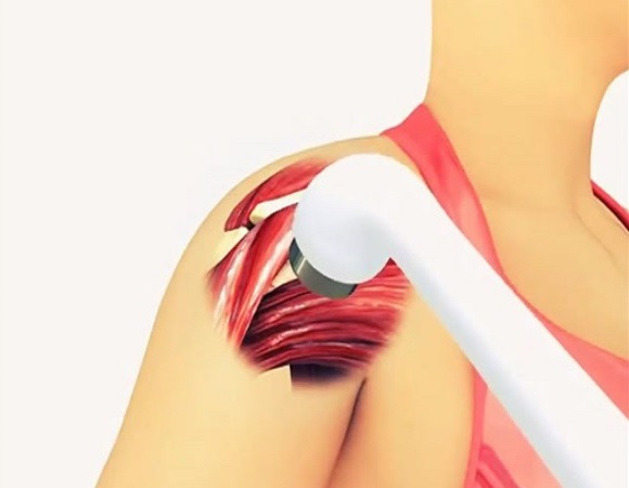
میکروماساژ مکانیکی
به هنگام فشردگی و انبساط محیط ، امواج طولی فراصوتی روی بافت اثر می گذارند و باعث جابجایی آب میان بافتی و در نتیجه باعث کاهش
ورم ( تجمع آب میان بافتی در اثر ضربه به یک محل) می شوند. درمان آسیب تازه و ورم : آسیب تازه معمولاً با ورم همراه است. فراصوت در
بسیاری از موارد برای از بین بردن مواد دفعی در اثر ضربه و کاهش خطر چسبندگی بافت ها به هم بکار می رود. درمان ورم کهنه یا مزمن:
فراصوت چسبندگی هایی که میان ساختمان های مجاور ممکن است ایجاد شود را می شکند.
خطرات اولتراسوند
1)سوختگی: اگر امواج پیوسته و در یک مکان بدون چرخش بکار روند، در بافت باعث سوختگی می شود و باید امواج حرکت داده شوند.
2)پارگی کروموزومی: استفاده دراز مدت از امواج اولتراسوند با شدت خیلی بالا پارگی در رشته دی ان ای (DNA) را نشان می دهد.
3)ایجاد حفره یا کاویتاسیون: یکی از عوامل کاهش انرژی امواج اولتراسوند هنگام گذشتن از بافت های بدن ایجاد حفره یا کاویتاسیون می باشد.
همه ی محلول ها شامل مقدار قابل ملاحظه ای حباب های گاز غیر قابل دیدن هستند و دامنه بزرگ نوسان های امواج اولتراسوند در داخل
محلول ها می تواند بر روی بافت ها تغییرات بیولوژیکی ایجاد کند ( پارگی در دیواره ی سلول ها و از هم گسستن مولکول های بزرگ )
مزایا و معایب اولتراسوند
General Ultrasound
Ultrasound imaging uses sound waves to produce pictures of the inside of the body. It is used to help diagnose the causes of pain, swelling and
infection in the body’s internal organs and to examine a baby in pregnant women and the brain and hips in infants. It’s also used to help guide
biopsies, diagnose heart conditions, and assess damage after a heart attack. Ultrasound is safe, noninvasive, and does not use ionizing radiation.
This procedure requires little to no special preparation. Your doctor will instruct you on how to prepare, including whether you should refrain from
eating or drinking beforehand. Leave jewelry at home and wear loose, comfortable clothing. You may be asked to wear a gown.
What is General Ultrasound Imaging?
Ultrasound is safe and painless, and produces pictures of the inside of the body using sound waves. Ultrasound imaging, also called ultrasound scanning or sonography, involves the use of a small transducer (probe) and ultrasound gel placed directly on the skin.
High-frequency sound waves are transmitted from the probe through the gel into the body. The transducer collects the sounds that bounce back and a computer then uses those sound waves to create an image.
Ultrasound examinations do not use ionizing radiation (as used in x-rays), thus there is no radiation exposure to the patient.
General Ultrasound
Because ultrasound images are captured in real-time, they can show the structure and movement of the body’s internal organs, as well as blood flowing through blood vessels.
Ultrasound imaging is a noninvasive medical test that helps physicians diagnose and treat medical conditions.
Conventional ultrasound displays the images in thin, flat sections of the body. Advancements in ultrasound technology include three-dimensional (3-D) ultrasound that formats the sound wave data into 3-D images.
A Doppler ultrasound study may be part of an ultrasound examination.
Doppler ultrasound, also called color Doppler ultrasonography, is a special ultrasound technique that allows the physician to see and evaluate blood flow through arteries and veins in the abdomen, arms, legs, neck and/or brain (in infants and children) or within various body organs such as the liver or kidneys.
There are three types of Doppler ultrasound:
-
Color Doppler uses a computer to convert Doppler measurements into an array of colors to show the speed and direction of blood flow through a blood vessel.
-
Power Doppler is a newer technique that is more sensitive than color Doppler and capable of providing greater detail of blood flow, especially when blood flow is little or minimal. Power Doppler, however, does not help the radiologist determine the direction of blood flow, which may be important in some situations.
-
Spectral Doppler displays blood flow measurements graphically, in terms of the distance traveled per unit of time, rather than as a color picture. It can also convert blood flow information into a distinctive sound that can be heard with every heartbeat.
What are some common uses of the procedure?
Ultrasound examinations can help to diagnose a variety of conditions and to assess organ damage following illness.
Ultrasound is used to help physicians evaluate symptoms such as:
-
pain
-
swelling
-
infection
Ultrasound is a useful way of examining many of the body’s internal organs, including but not limited to the:
-
heart and blood vessels, including the abdominal aorta and its major branches
-
liver
-
gallbladder
-
spleen
-
pancreas
-
kidneys
-
bladder
-
uterus, ovaries, and unborn child (fetus) in pregnant patients
-
eyes
-
thyroid and parathyroid glands
-
scrotum (testicles)
-
brain in infants
-
hips in infants
-
spine in infants
Ultrasound is also used to:
-
guide procedures such as needle biopsies, in which needles are used to sample cells from an abnormal area for laboratory testing.
-
image the breasts and guide biopsy of breast cancer (see the Ultrasound-Guided Breast Biopsy page.
-
diagnose a variety of heart conditions, including valve problems and congestive heart failure, and to assess damage after a heart attack. Ultrasound of the heart is commonly called an “echocardiogram” or “echo” for short.
Doppler ultrasound images can help the physician to see and evaluate:
-
blockages to blood flow (such as clots)
-
narrowing of vessels
-
tumors and congenital vascular malformations
-
reduced or absent blood flow to various organs
-
greater than normal blood flow to different areas, which is sometimes seen in infections