
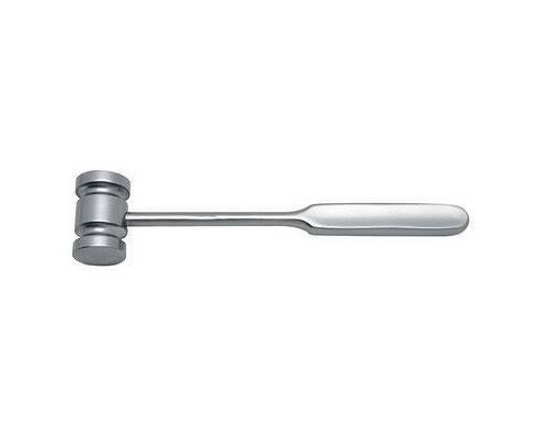
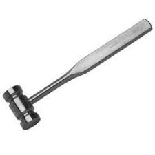
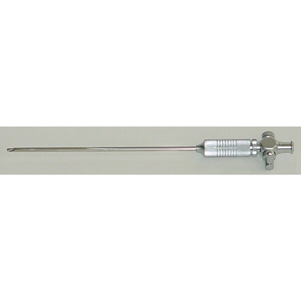
چکش ( Mallet )
نام وسیله : چکش ( Mallet )
چکش hammer : یک چکش از جنس استیل ضد زنگ ، یا استیل ضد زنگ پرشده با برنج است که وزن آن معمولا حدود 1-3 پوند است . ازچکش ها درسایر تخصص هایی که با استخوان سرکاردارند نیز استفاده می شود .
مورد استفاده : جهت جاگذاری یا خارج کردن ایمپلنت ها یا وارد آوردن نیرو به استئوتوم ها ،چیزل ، گوژها ،تامپ و سایر ابزارهای تخصصی به کارمی رود .
توضیحات بیشتر : به همراه استئوتوم ها ،چیزل ، گوژها ،تامپ و … استفاده می گردد .
برای مشاهده صفحه اینستاگرام مامایی می توانید بر روی این لینک کلیک نمائید .
برای مشاهده و خرید انواع ابزار های جراحی عمومی می توانید بر روی این لینک کلیک نمائید .
Mallet
Mallet
برای مشاهده محصولات دیگر برروی کلمه ی “ورودبه سایت“ کلیک نمایید./
-Click on the “Login to site“ to view other products. /
hammer
Soft – t issue i njuries and h ealing
The term soft tissues refers to those parts which are
not bone or cartilage. From the point of view of
injuries, it is necessary to consider the skin,
muscles, tendons, ligaments, blood vessels and
nerves (the latter are discussed in Chapter 3 ). It is
vital to consider not only what structures have
been damaged, but also how the damage has come
about, known as the mechanism of injury .
Mechanism of i njury
Injuries may be either blunt or penetrating. They
may occur by external insult, such as a cut by a
knife, or indirectly, such as a nerve damaged by the
sharp end of a bone. The amount of energy
imparted to the soft tissues is proportional to the
degree of violence applied.
چکش
Wound h ealing
Before discussing particular injuries, it is useful to
consider how tissues heal. Many tissues do not
regenerate when damaged and are repaired by collagenous scar tissue. Some tissues, such as bone,
mucous membrane, liver and the superfi cial layers
of skin, are capable of complete repair.
Wound h ealing p rocesses
The basic processes are seen best in the healing of a
clean incised wound:
1.
The wound bleeds and fi lls with clot
2.
The infl ammatory process is initiated and there
is dilatation of capillaries, exudation of fl uid and
white cells, and the process of capillary budding
begins
3.
Dead tissue and clot are removed by phagocytes,
and capillaries and fi broblasts migrate into the
damaged area. The new tissue is known as granulation tissue and is highly vascular (2 – 3 days)
4.
The skin surface begins to heal by the proliferation and migration of epithelial cells from the
edges of the wound to cover the defect
5.
The cellular reaction diminishes and the fi broblasts start to lay down collagen fi bres (third day
onwards)
6.
Vascularity diminishes and the collagen
increases
7.
Scar contraction makes the defect much smaller.
This effect is more marked in some areas than
others, e.g. in the midline of the body, particularly
over the back (2 weeks onwards)
8.
Scar consolidation and further shrinkage occurs,
and the scar becomes almost avascular.
The tensile strength of a wound increases to a
safe functional level in 15 days and is back to
normal in about 3 months, depending on the
tissue.
چکش
Closure of w ounds
With all wounds, particularly those communicating with a fracture or a joint, a decision has to be
made as soon as possible about closure. This decision will normally depend on the degree of contamination of the wound, the extent of surrounding
soft – tissue damage, the condition of the surrounding skin and the time which has elapsed since the
injury. The decision may be altered by the circumstances of follow – up. Where the patient can be kept
under observation, primary wound closure may be
attempted in circumstances which might be considered too risky if close follow – up were not possible as, for example, in wartime conditions. If the
decision has been made to close the wound, this
should be done as soon as possible. Closure is best
achieved with skin, either by direct suture or, for
larger defects, by skin grafting.
1.
Primary closure is usually safe if carried out in
the fi rst 6 hours after injury, provided all foreign
material and dead tissue is removed, there is no
communication with a fracture and there is little
surrounding soft – tissue damage. A clean incised
wound may be safely sutured up to 8 hours after
injury. After this time, contamination is almost
unavoidable and the risk of infection is much
greater. It is then usually safer to leave the wound
open and, after 24 hours, if it remains clean,
perform:
2.
Delayed primary closure .
3.
Secondary closure, which means closure after
the wound has been allowed to granulate, having
overcome any sepsis. This may be at 4 – 5 days or up
to several weeks after the injury. Suture may still be
possible at this late stage, but frequently, skin grafting will be necessary. Large defects fi ll initially with
granulation tissue, which is very resistant to infection. An area of clean granulation tissue is the best
bed for a skin graft when primary grafting is not
possible. Grafting in the presence of severe infection is usually unsuccessful, and tendons, ligaments
and, particularly, articular cartilage, do not usually
form suitable beds for non – vascularized grafts.
Following burns, areas of skin slough may need
to be excised when demarcation has occurred, and
the defect may then be covered by a suitable graft.
Techniques of c losure
چکش
Suture
Suture materials may be absorbable or non – absorbable. Sutures are either deep or superfi cial and may
be put in as a continuous stitch or as interrupted
individual stitches. Skin may alternatively be
apposed by adhesive dressings (e.g. butterfl y
sutures).
Skin g rafting
1.
Partial thickness or split – skin grafting. This is
the easiest and most reliable technique and uses
split skin taken from a convenient donor site. It
may be used as a primary or secondary technique.
It utilizes only part of the thickness of the epidermis and, if it is correctly taken, the donor site
should bleed from the skin papillae only and will
re – epithelialize spontaneously.
The graft may be held in place with dressings or
suture. ‘ Superglue ’has also been used with success
to hold grafts in place during healing.
2.
Full – thickness detached grafts. Until recently
these have been rarely used except for small areas,
such as defects on the fi ngers. When used as free
grafts, they are much less likely than split skin to
‘ take ’adequately. With the development of techniques for microvascular anastomosis, there is
now much more interest in using various types of
‘ free ’full – thickness grafts, either of skin alone or
using thicker grafts of skin, subcutaneous tissue
and muscle. It is also possible, for special requirements, to transfer composite grafts of skin, soft
tissues and bone, a technique which is being
increasingly used for the management of diffi cult
open fractures, particularly of the tibia.
3.
Attached skin fl aps. These are the more conventional types of full – thickness grafts which may
be rotated or swung, taken from one limb to
another, or from chest or abdomen to limb.
Considerable skill is needed to obtain good results.
They provide much more satisfactory skin cover,
but leave a defect elsewhere, which has to be closed
by split skin. They resist pressure better and are
essential for exposed and prominent areas. They are usually detached from the donor site in 10 – 15
days and may require further adjustment later.
4.
چکش
Foreign skin. Taken either from animal or
human, this is occasionally used as a temporary
dressing for large areas of loss, e.g. after burns. It is
eventually rejected and secondary grafting may
then be needed.
چکش


نقد و بررسیها
هنوز بررسیای ثبت نشده است.