
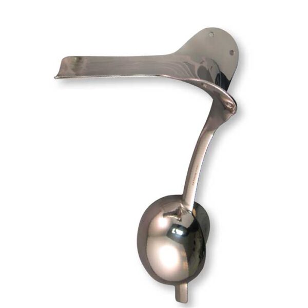
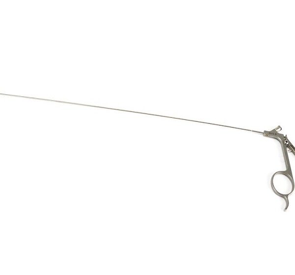
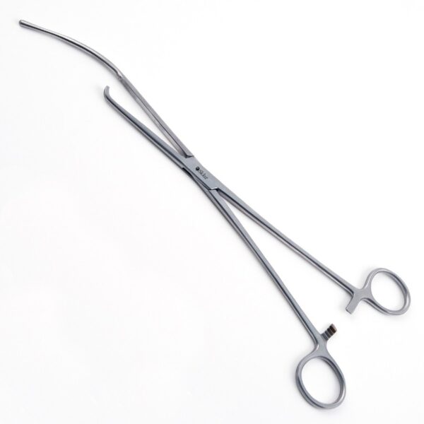
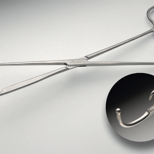
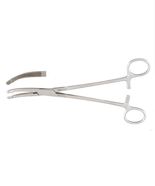
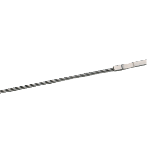
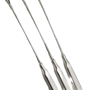
انبر خم کننده پلیت (PLATE BENDING PLIER )
انبر خم کننده پلیت (PLATE BENDING PLIER )
سایر اسامی : پلیت خم کن ( PLATE BENDER )
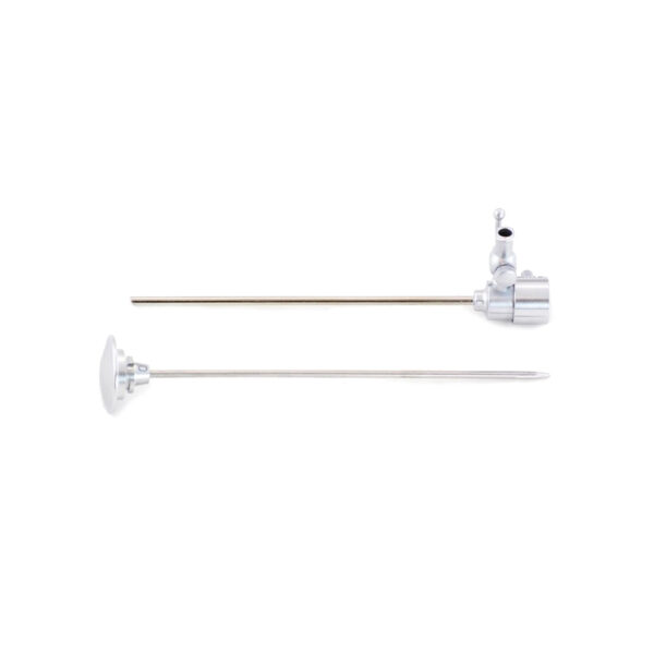
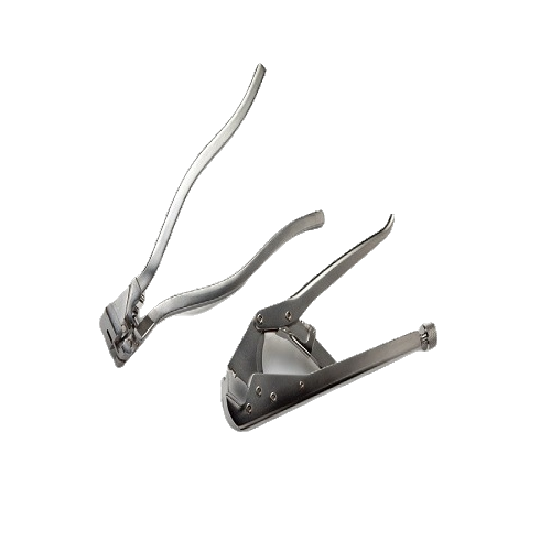
فورسپس های بزرگی هستند . پلیت مورد نطر را در بین آرواره های آن قرار داده و با فشردن دسته آن ، پلیت خم می گردد . در سایز ها و اشکال مختلف موجود است و بسته به نوع پلیت و سایز و نوع استخوان از آن ها استفاده می گردد .
مورد استفاده : در طول جراحی های فیکساسیون داخلی باز ف جهت خم کردن پلیت و شکل دهی آن جهت سازگاری با استخوان کاربرد دارد .
اغلب پلیت خم کن ها ، در ست های فیکساسیون موجود هستند .
برای مشاهده ی صفحه ی اینستاگرام ما می توانید بر روی این لینک کلیک نمایید.
(PLATE BENDING PLIER )
برای مشاهده محصولات دیگر برروی کلمه ی “ورودبه سایت“ کلیک نمایید./
–-Click on the “Login to site“ to view other products. /
A BRIEF INTRODUCTION INTO ORTHOPAEDIC IMPLANTS: SCREWS, PLATES, AND NAILS
Introduction:
Bone, as a hard biological tissue, consists of cells resided in the bone matrix, which is
made by an organic (primarily collagen (90%) and 10% amorphous ground substance) and a
mineral phase. The main constituents of bone mineral are calcium phosphate and calcium
carbonate. The mineral components consist mainly of hydroxyapatite crystals and amorphous
calcium phosphate. Bone acts as a reservoir for our body’s calcium, also serves as a protector
for vital organs, as well as provides mechanical stability to our body, and makes locomotion
possible. This hard biological tissue has a hierarchical structure and is designed optimally.
There are two types of bone, at the macroscopic level, in our body: cortical (compact or
Haversian) and cancellous (spongy or trabecular) bone (see Fig. 1). This specific
classification is based on the difference in their microstructure and basic units, as well as in
the extent of their porosities. Cortical bone’s porosities are at the micro-scales (the size of
voids are on the order of a couple of micrometers, and the porosity of cortical bone varies
from 5 to 30%), while cancellous bones’ porosities range from 30% to more than 90% [1].
Fig. 1. Cortical (compact or Haversian) and cancellous (spongy or trabecular) bone in femur (left) and vertebra
Based on the arrangements
انبر خم کننده پلیت
Based on the arrangements of the collagen fibrils, compact and spongy bones can be
classified either as woven or parallel-fibered type.
Woven bone, also called coarse-fiber bone,is characterized by the presence in the matrix of coarse, irregularly oriented collagen fibrils.
Woven bone is the bone formed during skeletal embryogenesis, and after birth it is gradually
removed by the process of bone remodeling, and is substituted by lamellar bone.
It should be noted that the woven bone be formed in pathological conditions such as callus formation, as well, It can be said that woven and lamellar bone are the result of a rapid and a slow
osteogenic process, respectively.
On the other hand, parallel-fibered bone consists of relatively thin and parallel-oriented collagen fibrils. Lamellar bone may be regularly organized into unit layers called lamellae.
Bone can gain maximum strength
Bone can gain maximum strength with minimum mass due to continuous activities of
various kinds of bone cells, i.e. osteocytes, osteoblasts, osteoclasts, and bone lining cells.
There are continuous processes of bone resorption and formation in our bones from birth to
death, which is so-called, bone remodeling process [4].
Bone remodeling process aims to provide maximum strength with minimum mass to our bones.
When a bone is broken, there is no other way than fixing it by employing man-made supportive structures.
Fortunately, bonehas a great capability in re-gaining its lost strength through the healing process.
The healing process of bone is a complex process in which both medicine and
mechanics are greatly at play and they can alter the time course of the healing process.
Healing process
Interesting to note that all broken bones go through the same healing process.
The bone healing process has three stages: inflammation, bone production (soft callus formation stage, and hard callus formation stage), and bone remodeling.
The inflammation stage begins the moment the bone is broken and lasts for around five days.
Fortunately bone has a very good blood supply due to the channels within its structure.
When a fracture occurs, there is massive disruption to these blood channels and a large amount of bleeding appears from the fracture fragments.
This is what causes immediate swelling and bruising in the area of the broken
bone. This is known as a Hematoma, which means bleeding within the tissue.
The damaged bone tissue at the edges of the fracture fragments die back and the dead cells release
chemicals called cytokines, which initiate the healing process.
Within hours of the fracture,the blood from the fracture fragments forms a mesh of clotted blood, which is the first linkbetween the two fragments and contains special cells called fibroblasts.
Fibroblasts begin to lay down tissue called granulation tissue between 4 and 10 days after the fracture occurs.
The granulation tissue forms a scaffold between the two fragments from which the formation of a
soft callus can begin.
انبر خم کننده پلیت
Fibroblast cells
Fibroblast cells present in the granulation tissue begin to form cartilage and fibrocartilage.
This is a spongy material that fills the gap between the two fracture fragments, although it is quite weak to external mechanical stimuli for around six weeks.
For this reason it is important that there is not too much movement of the fracture fragments at
this stage.
After a couple of weeks, despite the very fragile nature of soft callus, it offers sufficient stability at the fracture site for new blood vessels to begin forming and for osteoblasts at the periosteum (the outer surface of the bone) to begin laying down what is called woven bone.
This woven bone at the margins of the fracture is a little soft and disorganized, but it makes the first bone contact between the two fracture fragments.
From two to three weeks onwards a process begins whereby the fragile cartilage material of the soft callus is transformed completely into woven bone.
This process typically continues for between six and twelve weeks, depending on the location and type of fracture (generally six weeks for the upper limb and twelve weeks for the lower limb).
Hard Callus
Hard Callus formation is a complex process that is guided by the release of mineral compounds
such as Calcium and Phosphate into the Cartilage tissue, which subsequently transforms into a bridge of Hard Callus over the fracture site.
Once the hard callus has formed at the fracture site, then fracture union is said to have occurred.
Fracture union
Fracture union can be seen on x-ray at around six weeks in upper limb fractures and twelve weeks in lower limb fractures. During normal bone healing the body will lay down harder callus than is needed , and as a result the fracture site enlarges.
Bone remodeling begins once the fracture has united and may continue for several
years. Through the remodeling process and over time, the normal shape of the bone is
regained. Bone is resorbed where is not needed by osteoclasts and formed by osteoblasts
where is needed.
At the bone remodeling stage of fracture healing, a progression of weight bearing exercise is encouraged because it leads to an increase in bone strength.
انبر خم کننده پلیت
During the course of the bone remodeling process
During the course of the bone remodeling process, the loosely organized woven bone is gradually
replaced by lamellar bone, which is highly organized and much stronger than woven bone.
Orthopedic implants can be defined as medical devices used to replace or provide fixation of bone , or to replace articulating surfaces of a joint.
In simpler words, orthopedic implants are used to either assist or replace damaged or troubled bones and joints.
Orthopedic implants are mainly made from stainless steel and titanium alloys for strength and lined with plastic to act as artificial cartilage in order to reduce the stress at the articulating surfaces.
Some implants are cemented into place and others are pressed to fit, so that your bone can
grow into the implant for strength. Some examples of orthopaedic implants are: orthopaedic
plates, orthopaedic nails, and orthopaedic screws. The key factor that guides bone healing is
the interfragmentary movement, which determines the tissue strain and consequently the
cellular reaction in the fracture healing zone. Thus, the methods of fracture fixation will be
evaluated by considering their ability to reduce the interfragmentary movement. To achieve
good and acceptable healing results, biomechanical principles should be understood and
carefully taken into consideration.
انبر خم کننده پلیت

نقد و بررسیها
هنوز بررسیای ثبت نشده است.