
دفیبریلاتور (الکتروشوک)
دفیبریلاتور که با نام الکتروشوک شناخته می شود، دستگاهی است که فیبریلاسیون بطنی را از بین می برد.
فیبریلاسیون بطنی زمانی رخ می دهد که فیبرهای عضله قلب به صورت نامنظم منقبض می شود.
دفیبریلاتور
دفیبریلاتور جهت اصلاح این وضعیت به کار می رود. شوک اعمال شده به عضله قلب تمام فیبرهارا به طور
همزمان منقبض می کند و در نتیجه همه فیبرها به حالت اولیه می روند و امید است که بعد از آن به ریتم
طبیعی خود بازگردند.
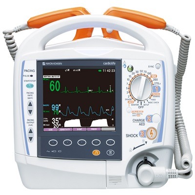
اصول عملکرد دفیبریلاتور
دفیبریلاتور، دستگاهی است که شوک الکتریکی را به عضله قلب میرساند. قبل از ۱۹۶۰، این سیستم به
صورت AC ارائه میشود که جریان ۵ تا ۶ آمپر یا فرکانس ۶۰ هرتز را در مدت ۲۵۰ تا ۱۰۰۰ میلی ثانیه در عرض
قفسه سینه بیمار اعمال میکردند. این روش عملاً موفقیت چندانی در اصلاحتارلرزه دهلیزی نداشت و اغلب
آریتمیهای جدیدتری ایجاد میکرد. دستگاههای امروزی که تکامل اساسی یافتهاند در زمان روشن بودن
دستگاه توسط خازنهای موجود در دستگاه، توانایی ذخیره ۴۰۰ ژول انرژی را دارا میباشند. از ۱۹۶۰ به بعد،
تارلرزهبرهای DC مختلف ساخته شد. این سیستمها شارژ dc را ذخیره میکند و روی بدن بیمار دشارژ
میشود. تفاوت عمده بین این دستگاهها، در شکل موج آنها است. متداولترین شکل موجها، موج wnها، تک
پالس، تاخیر مخروطی و ذوزنقهای است.
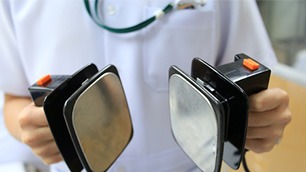
انواع دفیبریلاتور
دفیبریلاتورها پنج نوع هستند:
1. دفیبریلاتور دستی خارج از بدن Manual external
2. دفیبریلاتور دستی داخلی Manual internal
3. دفیبریلاتور خارجی اتوماتیک Automated external (AED)
4. کاردیوورتر_دفیبریلاتور قابل کاشت Implantable cardioverter-defibrillator (ICD)
5. دفیبریلاتور Wearable cardiac
دفیبریلاتور
Defibrillation
Defibrillation is a treatment for life-threatening cardiac dysrhythmias, specifically ventricular fibrillation (VF) and non-perfusing ventricular tachycardia (VT).A defibrillator delivers a dose of electric current (often called a countershock) to the heart. Although not fully understood, this would depolarize a large amount of the heart muscle, ending the dysrhythmia. Subsequently, the body’s natural pacemaker in the sinoatrial node of the heart is able to re-establish normal sinus rhythm.
In contrast to defibrillation, synchronized electrical cardioversion is an electrical shock delivered in synchrony to the cardiac cycle. Although the person may still be critically ill, cardioversion normally aims to end poorly perfusing cardiac dysrhythmias, such as supraventricular tachycardia.
Defibrillators can be external, transvenous, or implanted (implantable cardioverter-defibrillator), depending on the type of device used or needed. Some external .units, known as automated external defibrillators (AEDs), automate the diagnosis of treatable rhythms, meaning that lay responders or bystanders are able to use them successfully with little or no training.
Medical uses
Defibrillation is often an important step in cardiopulmonary resuscitation (CPR).CPR is an algorithm-based intervention aimed to restore cardiac and pulmonary function.Defibrillation is indicated only in certain types of cardiac dysrhythmias, specifically ventricular fibrillation (VF) and pulseless ventricular tachycardia. If the heart has completely stopped, as in asystole or pulseless electrical activity (PEA), defibrillation is not indicated. Defibrillation is also not indicated if the patient is conscious or has a pulse. Improperly given electrical shocks can cause dangerous dysrhythmias, such as ventricular fibrillation.
Survival rates for out-of-hospital cardiac arrests are poor, often less than 10% Outcome for in-hospital cardiac arrests are higher at 20%. Within the group of people presenting with cardiac arrest, the specific cardiac rhythm can significantly impact survival rates. Compared to people presenting with a non-shockable rhythm (such as asystole or PEA), people with a shockable rhythm (such as VF or pulseless ventricular tachycardia) have improved survival rates, ranging between 21-50%.
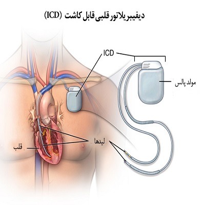
دفیبریلاتور قلبی قابل کاشت