

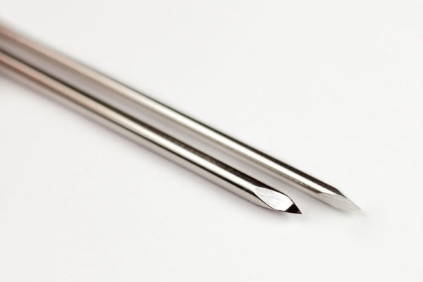
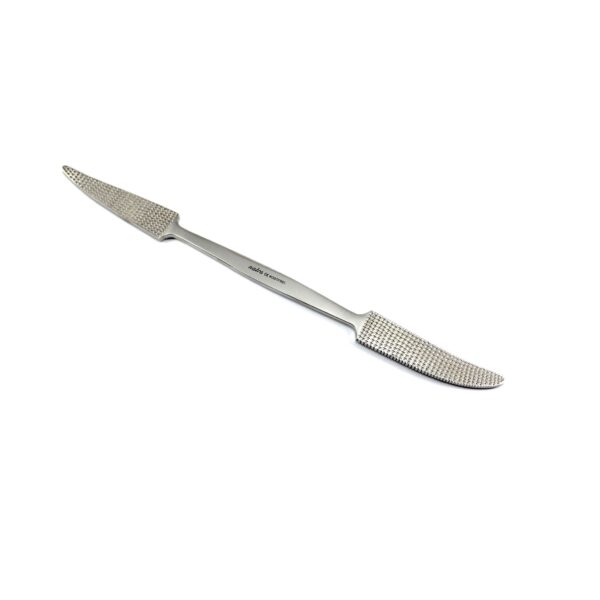
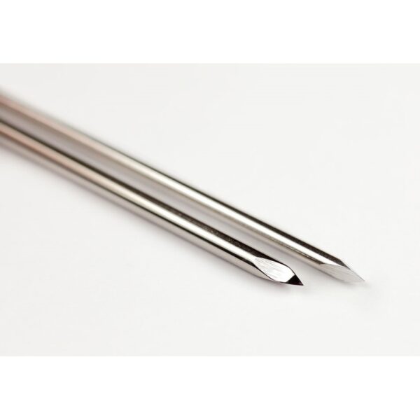
نام وسیله : پین استینمن صاف
smooth pin پین استینمن صاف ( smooth steinman pin ) :
این پین ها از جنس استیل ضد زنگ هستند که دارای یک نوک تروکار مانند لوزی شکل می باشد . این پین ها در سایز های 2-8/4 میلی متر ( 64/5 -16/3 اینچی ) موجود هستند .
مورد استفاده :
از این پین جهت ثابت کردن شکستگی های استخوانی ، تراکشن های اسکلتی ، ترمیم استخوانی و به عنوان یک گاید جهت گداشتن ایمپلنت ها مورد استفاده قرار می گیرد .
این نوع ازپین ، اغلب در استخوان های بزرگ کاربرد دارند .
توضیحات بیشتر :
در هنگام کار بااین وسیله باید مراقب بود زیرا نوک تیز آن می تواند به راحتی به پوست آسیب بزند .
برای مشاهده و خرید انواع ابزار های جراحی عمومی می توانید بر روی این لینک کلیک نمائید .
برای مشاهده صفحه اینستاگرام مامایی می توانید بر روی این لینک کلیک نمائید .
smooth pin
smooth steinman pin
برای مشاهده محصولات دیگر برروی کلمه ی “ورودبه سایت“ کلیک نمایید . /
-Click on the “Login to site“ to view other products . /
INTRAMEDULLARY FIXATIONIntramedullary fixation,
smooth steinman pin
with all of its attendant problems, is the most readily used system of internal fixation in smallanimals.(6,11,37)In many respects it is the least understood and least sophisticated method.
The techniques are adaptedfrom use in humans, and very little research or further development of intramedullary fixation has taken place in veterinaryorthopaedics.
(19) In contrast to human orthopaedics where the number of appliances is limited only by one’s imagination,
the devices used in veterinary medicine include only the Steinmann pin,
Kirschner wire, Rush pin, and Kuntscher nail, and ofthese, the Rush pin and Kuntscher nail are not used extensively.
STEINMANN PINNINGINDICATIONSIntramedullary
pinning with a single Steinmann pin may be indicated in fractures throughout the length of a long bone. It isbest for transverse and short oblique fractures of the middle third of long bones.
It can be applied in conjunction withcerclage and hemicerclage wiring, which will extend its indications considerably.
Single or multiple Steinmann pins togetherwith cerclage and hemicerclage wiring may be adapted for all types of fracture fixation.
BIOMECHANICST
smooth steinman pin
he Steinmann pin, while the most commonly used intramedullary device in veterinary medicine, is the least sophisticated.
Because it is placed in the medullary cavity,
it resists bending in all directions.
Its strength is related to its diameter, and itsability to restrict motion of the fracture fragment is related to its contact with the surrounding bony cortex.
A smallintramedullary pin in a large medullary cavity has been shown to be a successful method of producing nonunions (Fig. 16-1,A).
smooth steinman pin
Complications of intramedullary pinning with Steinmann pins can often be traced to mechanical factors such as pinmigration, bending, or catastrophic failure.
Delayed union and nonunion after intramedullary pinning may also be a result ofmechanical factors.
Since the medullary cavities of most bones in the dog vary widely in diameter, the Steinmann pin isusually used in three-point fixation.
It is anchored at the point of introduction,
smooth steinman pin
has contact with the fractured surfaces and/orthe isthmus of the medullary canal, and is impacted into the distal cancellous bone (Fig. 16-1, B).
A round intramedullary pincan do little to prevent torsional instability in fracture fixation.
The Steinmann pin may allow torsional stability only when itleads to interdigitation of the fracture fragments.
In general, when an intramedullary Steinmann pin is placed alone forreduction and stability of a fractured long bone,
it should contact as much of the medullary cortex as possible in order toprovide some torsional stability.
If intramedullary fixation does not present adequate stability against rotation,
either multiplepinning, cerclage wiring, hemicerclage wiring, or external fixation must be used in conjunction with the pinning.
Intramedullary devices provide no longitudinal support;
the fracture fixation is dependent upon the stability of the fracturefragments themselves.
Therefore, when comminution or cracks exist, there is a definite possibility for further collapse andconverted by Web2PDFConvert.com
telescoping of the fracture fragments over the Steinmann pin.
FIG. 16-1
Intramedullary pin size compared with medullary cavity diameter.
پین استینمن صاف
(A) A smallintramedullary pin allows significant movement and may result in pin loosening or nonunion.
(B)A larger pin restricts movement and therefore aids healing.
This drawing shows a properlyfitting Steinmann pin that is anchored at three levels proximal femur, isthmus at fracture site,and distally in the soft cancellous bone of the metaphysis.
METHOD OF INSERTIONT
smooth steinman pin
he Steinmann pin is inserted by using a Jacob’s chuck.
The pin should be introduced at the end of the bone, cross thefracture site, and become embedded in the distal metaphysis of the bone.
Retrograde insertion of Steinmann pins isadvocated by some and has the advantage of being technically easier to accomplish.
However, complications ofinadequate positioning and soft tissue (usually nerve) damage can be caused by improper retrograde pinning.
The site forintroducing a Steinmann pin in the dog’s femur is in the subtrochanteric fossa.
It may also be introduced distally in the femurthrough the intercondylar fossa (Fig. 16-2).
smooth pin
In the humerus it is introduced through the proximal lateral aspect.
The point ofintroduction is determined by the curvature of the humerus of the individual patient.
In the humerus the Steinmann pin isstarted as far proximally as possible to still allow the pin to be positioned distally at the medial distal epicondyle.
In the tibia,the Steinmann pin is introduced just medial and slightly behind the straight patellar ligament.
smooth steinman pin
In this position the pin entersthe medullary cavity in front of the joint without invading it.
In the ulna the Steinmann pin is inserted through the olecranonparallel to the medullary cavity of the ulna.
Sometimes the intramedullary pin may be introduced retrograde in the ulna, but itis important that its direction be characterized so that the pin does not penetrate the elbow joint.
I do not recommendintramedullary pinning of the radius owing to the inability to adequately stabilize the radius without interfering with a jointsurface.
Although introduction of the Steinmann pin into the radius in a retrograde fashion passing it out through the distalradiocarpal joint has been advocated in the past, there are better methods to achieve fixation of this bone.
FIG. 16-2 Pin position is shown for insertion through the intercondylar fossa of the distal femurin the treatment of supracondylar fractures.
The seating of a Steinmann pin is a very important part of its placement.
smooth pin
After the pin has been introduced, crossed thefracture site, and reduced the fracture, it should be seated firmly in the distal fragment.
The distance traveled by the pin issometimes difficult to measure and should be planned before introduction into the distal fragment.
This can be done easilyby adjusting the chuck on the pin so that an adequate amount of pin will appear between the chuck and the surface of theskin;
this distance is equivalent to the length of the pin that needs to be introduced into the distal fragment.
steinman pin
Using thistechnique, it is relatively easy to establish a firm seating for the Steinmann pin without penetrating the joint surface below thefracture.
As seating of the pin is achieved,
the rotation that is necessary to insert the pin should be lessened to the pointwhere only longitudinal pressure is applied to push the pin so that it may interdigitate with the cancellous bone.
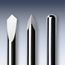
Steinmannpins are available with threaded tips.
smooth steinman pin
These threaded tips have no advantage and, in fact, cause stress concentration tooccur at the junction of the shaft and thread,
sometimes leading to breakage of the device.
The trocar point is most oftenused for drilling and usually allows adequate seating of the pin in the distal aspect of the bone.
steinman pin پین استیننمن صاف
Spade tips can be usedwhen very soft bone is found and the Steinmann pin can be seated with a mallet to prevent rotation of this pin in the bone.
After the pin has been cut off below the level of the skin,
final seating with a mallet and countersink may improve thepurchase of the pin in the cancellous bone of the metaphysis.
smooth pin
FOLLOW-UP TREATMENTFracture union is usually complete within 8 to 12 weeks following intramedullary pinning.
In younger animals,
this time maybe reduced, and it is necessary to follow the dog very closely, especially if he is a giant breed, so that the pin may beremoved before the bone “swallows” it up by growth.
Intramedullary pins of the Steinmann type are generally removed afterradiographs show evidence of periosteal bridging and usually after some degree of bone remodeling is noted in the callusitself.
In other words, the callus should be resorbing and not proliferating at the time that the intramedullary pin is removed.
steinman pin
پین استینمن صاف
Since the round intramedullary pin does not hold the fracture completely stable, considerable periosteal callus is sometimesnoted.
This callus is probably related to motion and will form a strong bridge across the fracture fragments, usuallyproducing a desirable end result.
The removal of the Steinmann pin is accomplished with a pair of
pliers, pin puller, orgrasping forceps by means of a stab incision through the skin.
Early postoperative immobilization of the animal isnecessary to prevent collapse or rotation of the fracture fragments following Steinmann pinning.
It is important that theamount of rotational stability be accurately determined at the time of surgery,
since further immobilization through the use ofcerclage and hemicerclage wiring or external splinting or casting techniques may be helpful in limiting this rotation.
Althoughevery attempt is made to immobilize the animal, active movement of the joints and muscles of the affected extremity isencouraged.
CONTRAINDICATIONS AND COMPLICATIONS
پین استینمن صاف
smooth pin
Single Steinmann pins are usually contraindicated in severely comminuted fractures except when cerclage or hemicerclagewiring is added.
Intramedullary pins are not good devices to use in the presence of sepsis.
Insertion of a Steinmann pin intothe marrow cavity of a septic fracture may cause the extension of infection throughout the medullary cavity and make thetreatment of this infection very difficult.
Steinmann pinning should be attempted only when the fracture can be made stable.
smooth pin
Inadequate technique of Steinmann pin placement with improper seating in the distal fragment or instability of the fracturewill lead to the complication of pin migration,
the most common problem associated with intramedullary pinning.
It is seen inanimals where there is instability at the fracture site,
allowing the fracture to collapse over the pin, or where there is sufficientmotion to cause loosening of the pin at its distal aspect.
If the pin loosens, the fracture will usually distract or collapse andconverted by Web2PDFConvert.com
smooth steinman pin
angulate.
The pin may penetrate the skin through the site of initial insertion and create a tract for infection.
The Steinmannpin should never be allowed to protrude through the skin when it is used for an intramedullary function.
If this occurs, the pinshould be removed and replaced immediately with a larger pin or some other form of fixation that will adequately stabilizethe fracture.
smooth pin پین استینمن صاف
Simply reinserting the same pin after it has penetrated through the skin is not adequate treatment; this mayserve only to be the focus for an infectious process, and the pin will usually begin backing out again several days later.
If pinmigration is a problem, it is evidence of instability at the fracture site and should be corrected immediately.
Instability of thefracture, especially of the femur, will allow the formation of rotational deformities,
usually an external rotation of the proximalfragment as a result of the pull of the iliopsoas muscle.
This complication is very common and can be seen on the lateralradiograph (Fig. 16-3), which shows a femoral head that faces cranially.
It will produce an abnormality in gait and should beprevented.
smooth pin
smooth steinman pin
seo & apload 0to100 by M.F

نقد و بررسیها
هنوز بررسیای ثبت نشده است.