
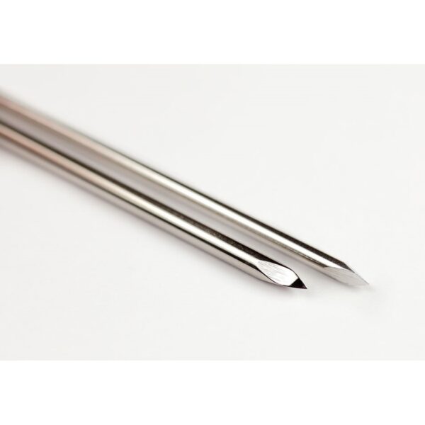
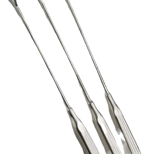
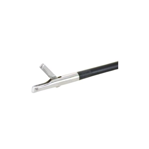
نام وسیله : پین استینمن دنده دار
THREADED PiNS پین استینمن دنده دار ( THREADED steinman pins ) :
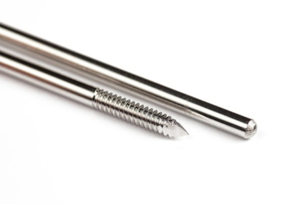
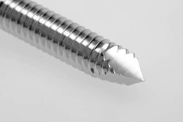
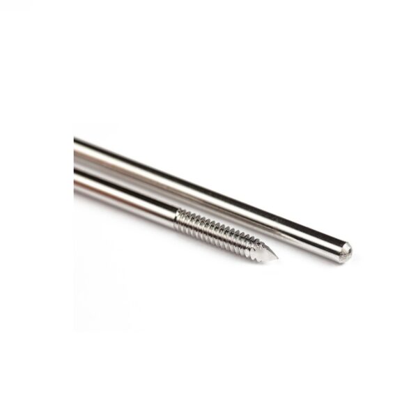
این پین ها از جنس استیل ضد زنگ هستند که دارای یک نوک تروکار مانند دنده دار می باشد . این پین ها در سایز های 2-8/4 میلی متر ( 64/5 -16/3 اینچی ) موجود هستند .
پین استینمن دنده دار
برای مشاهده محصولات دیگر برروی کلمه ی “ورودبه سایت“ کلیک نمایید . /
-Click on the “Login to site“ to view other products . /
مورد استفاده :
از این پین جهت ثابت کردن شکستگی های استخوانی ، تراکشن های اسکلتی ، ترمیم استخوانی و به عنوان یک گاید جهت گداشتن ایمپلنت ها مورد استفاده قرار می گیرد .
این نوع ازپین ، اغلب در استخوان های بزرگ کاربرد دارند .
threaded steinmann pin
توضیحات بیشتر : در هنگام کار بااین وسیله باید مراقب بود زیرا نوک تیز آن می تواند به راحتی به پوست آسیب بزند .
برای مشاهده و خرید انواع ابزار های جراحی عمومی می توانید بر روی این لینک کلیک نمائید .
، ، ، ، ، ، ، ، ، ، ، ، ، ، ، ، ، ، ، ، ، ، ، ، ، ، ، ، ، ، ، ، ، ، ، ، ، ، ، ، ،، ، ، ، ، ، ، ، ، ، ، ، ، ، ، ، ، ، ، ، ، ، ، ، ، ، ، ، ، ، ، ، ، ، ، ، ، ، ، ، ،
برای مشاهده صفحه اینستاگرام مامایی می توانید بر روی این لینک کلیک نمائید .
The current study was undertaken to evaluate the clinical efficacy of end-threaded intramedullary pinning for management of various long bone fractures in canines.
Materials and Methods:
This study was conducted in two phases, managing 25 client-owned dogs presented with different fractures.
The technique of application of end-threaded intramedullary pinning in long bone fractures was initially standardized in 6 clinical patients presented with long bone fractures.
In this phase, end-threaded pins of different profiles, i.e.,positive and negative, were used as the internal fixation technique.
On the basis of results obtained from standardizationphase, 19 client-owned dogs clinically presented with different fractures were implanted with end-threaded intramedullary positive profile screw ended self-tapping pin in the clinical application phase.
.
THREADED PiNS پین استینمن دنده دار
Results:
The patients, allocated randomly in two groups, when evaluated postoperatively revealed slight pin migration in Group-I (negative profile), which resulted in disruption of callus site causing delayed union in one case and large callus formation in other two cases whereas no pin migration was observed in Group-II (positive profile).
Other observations in Group-I was reduced muscle girth and delayed healing time as compared to Group-II.
In clinical application, phase 21stand 42ndday post-operative radiographic follow-up revealed no pin migration in any of the cases, and there was no bone shortening or fragment collapse in end-threaded intramedullary positive profile screw ended self-tapping pin.
THREADED PiNS
Conclusion:
The end-threaded intramedullary positive profile screw ended self-tapping pin used for fixation of long bone fractures in canines can resist pin migration, pin breakage, and all loads acting on the bone, i.e.,compression, tension, bending, rotation, and shearing to an extent with no post-operative complications.
THREADED PiNS
Keywords:
Admit pin, canine, end-threaded, fracture, intramedullary, orthopedics, pinning, positive profile.IntroductionManagement of fractures through intramedullary fixation is regularly used in Veterinary Orthopaedics.
The Steinmann pin appears to be the most often used material, either on its own or in combination, while the Rush pin, Kirschner wire, Kuntscher pin, and interlocking pin have all been employed depending on their indication.
Unthreaded intramedullary pins alone cannot provide adequate traction and rota-tional stability, as they are weak against rotational and shearing forces [1].
Stack pin application par-tially prevents these disadvantages by opposing the horizontal crossing and bending forces [2], and it has been reported that combined plate-intramedullary pin application is successful in increasing axial and rota-tional stability[3].
Intramedullary interlocking nailing is used to achieve rigid repair which can counteract all forces and are entirely load bearing until callus forma-tion[4].
Rotational stability can also be increased by cerclage wire, external fixation, interlocking pins, and trilam nails [5], or using a C-clamp on the plate [6,7] reported that stabilization of a Salter-Harris TypeIV physeal fracture of the humeral condyle in a miniature pinscher was simplified by using Orthofix partially threaded Kirschner wire, with excellent clinical results.
Partially threaded pins, having a negative profile end-ing create a weak point in the pin-thread junction, so if these pins are to be used, the junction must not be near the fracture line [8].
The pros and cons of vari-ous implants were taken into account and the innova-tion in terms of
“end-threaded intramedullary positive profile screw ended self-tapping pin” (Admit pin) was conceived to minimize the complications of frequently used economic routine intramedullary pinning.
The aim of this study was to standardize the tech-nique of application of end-threaded intramedullary.
pin for management of long bone fractures in canines and to evaluate the efficacy of end-threaded intramed-ullary pin in the management of long bone fractures in canines.
Materials and MethodsEthical approvalsThe research design is purely an applied clinical study, therefore the ethical approval from Institutional animal ethics committee was not mandatory.
However, the broad outline of the work has been approved by the committee.
پین استیننمن دنده دار
Design and manufacturing of Admit pinIntramedullary end-threaded pins (Figure-1)
were manufactured from an iron based alloy-316L Stainless Steel, in an end-threaded fashion.
Pins were produced in various diameters, ranging from 4.5 mm (major diameter)/4 mm (pitch diameter) to 7.5 mm/5.5 mm with a standard length of 9 inches.
The distal end of the pin was designed with a positive profile self-tap-ping screw pointed end (Figure-2)
to allow for ease of entry into the cancellous bone whereas the proximal end is kept trocared so that there is no need of
a pilot hole in proximal fragment of bone when used either in retrograde or normograde fashion (Figure-2).
Clinical application phaseIn the present study, 25 client-owned dogs of dif-ferent breeds, sex, and age presented in the Teaching Veterinary Clinical Complex of
DGCN College of Veterinary and Animal Sciences, Palampur, with various long bone fractures were treated under two phases.
The study was based on clinical cases, so there is no need of ethical approval as the cases were treated as per highest standard of the treatment at par with any national or international standards without harming or giving any unnecessary stress to the animals.
All patients were subjected to clinical and radiological examinations preoperatively, those required mild sedation for pre-operative evaluation were administered acepromazine at 0.05 mg/kg B.Wt, I.M.; butorphanol tartrate at 0.2 mg/kg B.Wt, I.M. and atropine sulfate at 0.04 mg/kg B.Wt., S.C.
The patient scheduled for surgery was given a standard protocol comprising injection butorphanol at 0.2 mg/kg B.Wt, I.V. and injection diazepam at 0.5 mg/kg B.Wt, I.V.
followed by injection propofol (till effect, I.V.) and maintenance by isoflurane throughout the period of surgery.
THREADED PiNS
The diameter and length of the implant were determined by pre-operative radiographs, size and weight of the dog and intra-operative assessment.
An open reduction with a lateral approach was the tech-nique of choice for diaphyseal, epiphyseal, or metaph-yseal femoral fracture repair;
craniolateral approach was preferred for humeral fracture repair, and medial approach was preferred for tibial fracture repair.
threaded steinmann pin
Research designIn
the first phase, 6 dogs were used for tech-nique standardization.
These six client-owned dogs
were randomly allocated into two groups.
In Group-I, three dogs with long bone fracture
(8, 3, and 1 years weighing 26 kg, 18 kg, and 20 kg, respectively)
were implanted with end-threaded intramedullary negative profile pin (Figure-3) with a trocar end of
5.0/5.2 mm, Whereas in Group-II, three dogs with long bone frac-tures
(18 months, 3 years, and 10 years weighing 16 kg, 20.5 kg, and 30 kg, respectively)
were implanted with end-threaded intramedullary positive profile pin screw ended self-tapping pin of 4.5/6.5 mm (Figure-3).
On the basis of the results obtained from Phase-I, 19 client-owned dogs
(age; 1.5 month – 9 years and body weight; 3.40-21 kg),
clinically presented with fracture of different long bones
(femur, humerus, and tibia)
پین استینمن دنده دار
were implanted with end-threaded positive profile intramedullary pin with a screw end of size:
3.5/4.0 mm, 4.5/5.0 mm, and 4.5/6.5 mm.ResultsIn standardization phase, pins were removed after complete attainment of weight bearing by the animals with proper radiographic evidence of frac-ture healing in both groups.
Evaluation at 21st and 42nd day postoperatively revealed slight pin migration in Group-I, which resulted in disruption of callus site causing delayed union in one case and large callus formation in other two cases whereas no pin migra-
tion was observed in Group-II.
Other observations in
Group-I was reduced muscle girth and delayed heal-
ing time as compared to Group-II. Rotational forces
were not resisted by the implant used in Group-I. On
the other hand, the implant used in Group-II success-
fully resisted rotational forces as the threads were
embedded in distal cancellous bone firmly (Table-1).
Partially threaded pins having a negative profile end-
ing create a weak point in the pin-thread junction, so
if these pins are to be used, the junction must not be
near the fracture line.
threaded steinmann pin
Positive profile pins do not have
this problem because the threads are raised above the
core diameter of the pin.
Thus, there is no stress riser
(weak point) at the thread non-thread interface and the
implant appears very sturdy (Table-1).
پین استینمن دنده دار
In clinical application phase, 19 cases were
studied (Table-2).
The parameters evaluated, 21st and
42nd day postoperatively showed an initial decrease in
muscle girth due to; removal of hematoma formed at
the site, reduction of fractured bone fragments back to
their normal position and post-operative resolution of
inflammatory process.
threaded steinmann pin
The gradual increase in mus-
cle girth observed over a period of time was due to;
increased blood supply to the site of fracture, regener-
ation of healthy muscle tissue and callus formation at
the fracture site.
threaded steinmann pin
نقد و بررسیها
هنوز بررسیای ثبت نشده است.