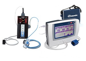
دستگاه کاپنوگراف (Capnograph)
دستگاه کاپنـــــوگراف از دو کلمه Capno به معنی دی اکسید کربن و Graph به معنی صفحه ی نمایش جهت
خواندن و ثبت کردن تشکیل شده است. این دستگاه میزان گاز CO2 در حجم هوای اشبـــاع شده از ریه های
بیمار را نشان می دهد.سلول های بدن انسان نیاز به اکسیژن دارند ، و این اکسیژن توسط خون به آن ها می
رسد. O2 به سلول ها تحویل داده می شود و پس از مصرف آن به وسیله ی سلــول ها، گازCO2 سمی تولید
شده با گردش خون خارج می شود. خون، گاز CO2 را به سمت مویرگ های موجود در ریه ها هدایت کرده تا در
بازدم های بعدی به فضای خارج فرستاده شوند.مانیتورهای دی اکسیـــد کربن فشار جزئی این گاز را در بازدم
بیمار اندازه گیری می کنند. معمولاً در پایان بازدم غلظت دی اکسیـد کربن به حداکثر مقدار خود می رسد، لذا
غلظت اندازه گیری شده در این حالت به دی اکسید کربن انتهای بازدمی معروف است.اتاق های عمـل اصلی
ترین محل به کار گیری این وسیله است.در زمان بیهوشی بیمار کاپنـــوگراف اطلاعات مفیدی در مورد وضعیت
تنفسی بیمــار ، قطع شدن مسیـر ونتیلاسیـــون و نشت هوای دمی به کادر پزشک می دهد. کاپنوگراف دی
اکسید کربن را در هنگام هر سیکل دم/بازدم اندازه گیری کرده وشکل موج آن را به همراه مقدار عددی نشـان
می دهند. مانیتورینگ های دی اکسید کربن غلظت این گاز را یا با استفاده از یک حسگر که به طور مستقیم در
مسیـر تنفس بیمار قرار دارند ( روش جریان اصلی ) اندازه گیـــری می کنند و یا در محلی دیگر با نمونه گیری از
مسیر هوایی بیمار ( جریان جانبی )
انواع کاپنوگراف
1-کاپنوگراف Main stream : دارای یک سنسور CO2 هستند، که به یک تعدیل کننده راه هوایی متصل شده
است. نور مادون قرمز توسط CO2 در جریان هوای بیرون رونده جذب می شود. هرچه میزان گاز CO2 بیشتر
باشد، میزان نور مادون قرمز بیشتری جذب شده و نور کمتری به آشکار ساز می رسد.
2-کاپنوگراف نوع Side stream : از گاز موجود در مسیر هوایی از طریق یک لوله کوچک نمونه برداری می کنند
در مکانی مشابه با محل قرار گیری سنسور کاپنوگراف Main stram تمامی اندازه گیری ها و پردازش سیگنال
در درون خود کاپنوگراف Side stream انجام می شود به روشی مشابه کاپنوگراف Main

دستگاه کاپنوگراف
Capnography
Capnography is the monitoring of the concentration or partial pressure of carbon dioxide (CO2) in
the respiratory gases. Its main development has been as a monitoring tool for use
during anesthesia and intensive care.It is usually presented as a graph of expiratory CO2 (measured
in millimeters of mercury, “mmHg”) plotted against time, or, less commonly, but more usefully,
expired volume.The plot may also show the inspired CO2, which is of interest
when rebreathing systems are being used.The capnogram is a direct monitor of the inhaled and
exhaled concentration or partial pressure of CO2, and an indirect monitor of the CO2 partial pressure
in the arterial blood. In healthy individuals, the difference between arterial blood and expired
gas CO2partial pressures is very small.In the presence of most forms of lung disease, and some
forms of congenital heart disease (the cyanotic lesions) the difference between arterial blood and
expired gas increases and can exceed 1 kPa.
Working mechanism
Capnographs usually work on the principle that CO2 absorbs infrared radiation.A beam of infrared
light is passed across the gas sample to fall on a sensor. The presence of CO2 in the gas leads to a
reduction in the amount of light falling on the sensor, which changes the voltage in a circuit. The
analysis is rapid and accurate, but the presence of nitrous oxide in the gas mix changes the infrared
absorption via the phenomenon of collision broadening.This must be corrected for measuring
the CO2 in human breath by measuring its infrared absorptive power. This was established as a
reliable technique by John Tyndall in 1864, though 19th and early 20th century devices were too
cumbersome for everyday clinical use.
Diagnostic usage
Capnography provides information about CO2 production, pulmonary (lung)
perfusion, alveolar ventilation, respiratory patterns, and elimination of CO2 from the anesthesia
breathing circuit and ventilator.The shape of the curve is affected by some forms of lung disease; in
general there are obstructive conditions such as bronchitis, emphysema and asthma, in which the
mixing of gases within the lung is affected.Conditions such as pulmonary embolism and congenital
heart disease, which affect perfusion of the lung, do not, in themselves, affect the shape of the
curve, but greatly affect the relationship between expired CO2 and arterial blood CO2. Capnography
can also be used to measure carbon dioxide production, a measure
of metabolism.Increased CO2 production is seen during fever and shivering. Reduced production is
seen during anesthesia and hypothermia.
Use in anaesthesia


