دستگاه فیکو
دستگاه فیکو توسط به کار گیری امواج اولتراسوند قادر است بخش عدسی چشم را جهت عمل آب مروارید برداشته و توسط ساکشن های مخصوصی خارج کند.
مکانیزم ایجاد کننده ی اولتراسوند در هند پیس فیکو سبب می شود که نوک متصل به آن با سرعت به سمت جلو و عقب نوسان کند. از قطعات اصلی ماشین یا دستگاه فیکو می توان به موارد زیر اشاره کرد:
رایانه :
شامل سخت افزار، صفحه ی نمایشگرو تنظیمات است که معمولا به صورت لمسی اداره می شود و در آن می توان مدهای مختلف ، تغییر پارامترهای مربوط
به قدرت فیکو، شدت مکش و سرعت برقراری چریان مایع را تنظیم نمود.
پمپ:
وکیوم نیروی مکشی است که توسط پمپ در نوک ایجاد می شود.
هند پیس پروبی است که به وسیله ی آن نیروی حاصل از امواج فراصوتی اعمال می شود.
کاست :
مکش به کاست اعمال می شود و لنز خرد شده و مایعی که در چشم جریان پیدا کرده به داخل کاست کشیده می شود. در صورتی که کاست
پر شده باشدمکش دچار توقف می شود. بنابراین لازم است وضعیت کاست توسط کمک جراح پایش شود و به هنگام ، کاست را جهت ادامه
عمل تخلیه نمود.
پدال فیکو:
جراح عملکرد های سه گانه هند پیس با استفاده از پدال کنترل می کند. برای پدال چهار موقعیت پلکانی 1،2،3،0 ذکر می شود.
فیکو
دستگاه فیکو
جراحی آب مروارید (فیکو)
کاتاراکت یا آب مروارید، یک بیماری چشمی است که در آن، ناحیه ابرمانند بدون دردی روی عدسی چشم ایجاد میشود. این ضایعه درواقع کدورت عدسی بیمار است که بصورت مهآلود شدن دید، خود را نشان میدهد. این حالت موجب اختلالاتی در دید و تاری دید میشود. آب مروارید اغلب در افراد مسن اتفاق میافتد. تصویر سمت چپ چشم سالم و تصویر سمت راست نشانگر یک آب مروارید کاملاً رسیده است.
درمان آب مروارید
علت آب مروارید
عدسی چشم ترکیبی از آب و پروتئین است. پروتئینها به نحوی قرار گرفتهاند که عدسی را شفاف نگه میدارند و باعث عبور نور از آن میشوند. آب مروارید به دنبال تغییراتی در ساختار پروتئینی عدسی چشم و تجمع آنها در بعضی نواحی عدسی ایجاد میشود. این حالت باعث به وجود آمدن ناحیهای کدر روی عدسی چشم میگردد. گرچه علت اصلی ایجاد آب مروارید مشخص نیست اما براساس عوامل ایجاد کننده، بیماری آب مروارید به چند دسته تقسیم میشود که در ادامه آورده شده است.
انواع آب مروارید
آب مروارید وابسته به سن: این نوع از آب مروارید که شایعترین علت است، در نتیجه افزایش سن ایجاد میشود.
آب مروارید مادرزادی: گاهی در نتیجه عفونت داخل رحمی، آسیب یا عدم تکامل کافی قبل از تولد، نوزادان مبتلا به آب مروارید به دنیا میآیند یا ممکن است در اوایل کودکی مبتلا به آب مروارید شوند.
آب مروارید ثانویه: در نتیجه برخی بیماریها مثل دیابت، سموم، مصرف داروهای خاص مثل کورتیکواستروئید یا دیورتیک و اشعه ماورای بنفش، آب مروارید میتواند ایجاد شود.
آب مروارید پس از ضربه: این نوع از آب مروارید به دنبال ضربه به چشم ایجاد میشود.
به طور کلی عواملی مثل دود سیگار، آلودگی هوا و مصرف زیاد الکل نیز میتوانند خطر ابتلا به آب مروارید را افزایش دهند.
برای اطلاعات بیشتر به این پیج مراجعه فرمایید .
What is Phacoemulsification
Definition:
Phacoemulsification is the most common cataract surgery technique performed. Cataract surgery is
used to restore vision in patients whose vision has become cloudy from cataracts, a clouding of the
eye’s lens.
The lens is located behind the iris. It is responsible for focusing light on the retina, and for producing
clear, sharp images. The lens has the ability to change shape, known as accommodation.
As the eyes age, however, the lens hardens and loses its ability to accommodate. The entire lens is
contained within a lens capsule. As the eyes age, oxidative processes occur and dead cells
accumulate in the lens capsule, causing the lens to gradually become cloudy. The light that would
normally be focused by the lens is scattered around because of the cloudiness, so vision is no longer
clear and sharp.
How is phacoemulsification performed?
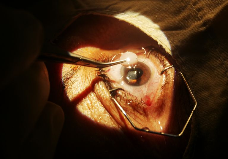
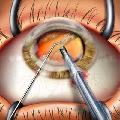
During phacoemulsification, a surgeon makes a small incision at the edge of the cornea and then
creates an opening in the membrane that surrounds the lens. A small ultrasonic probe is then
inserted, breaking up the cloudy lens into tiny fragments. The instrument vibrates at ultrasonic
speed to chop and almost dissolve the lens material into tiny fragments. The fragments are then
suctioned out of the capsule by an attachment on the probe tip.
After the lens particles are removed, an intraocular lens implant, commonly referred to as an IOL, is
implanted and positioned into the lenses natural capsule.
It is inserted through the tiny corneal incision through a hollowed out tube. Once the lens is pushed
through, it unfolds and is positioned in place.
Phacoemulsification is typically performed in an outpatient surgery center and normally does not
require a hospital stay. The cataract surgery procedure is performed under local anesthesia (an
anesthetic injected around the eye) or topical anesthesia (numbing drops inserted into the eye).
What is the recovery time for phacoemulsification?
The incision made in the cornea usually requires no stitches and is self-sealing. Within a few days,
the incision heals completely. Post-operative eye drops are prescribed and usually consist of
antibiotics, steroids, and a non-steroidal anti-inflammatory medication. These drops reduce
inflammation and prevent infection. The antibiotic is usually discontinued within 7-10 days. The
steroid and non-steroidal anti-inflammatory are taped over 3-6 weeks depending on the surgery.
Most patients have vision improvement almost immediately and vision tends to steadily improve over
4-5 weeks.
Phacoemulsification revolutionized cataract surgery. Before phacoemulsification was developed,
surgeons would remove the entire lens and capsule. This made it difficult to insert an intraocular
lens. The lens of the eye contributes a lot of focusing power to the eye. As a result, if you remove
the cataract, which is the lens, the patient is left with a very high “plus,” farsighted prescription. This
is why, many years ago, when patients had cataracts removed, they typically wore “cataract
glasses.” Cataract glasses were thick, heavy and magnified the eyes.
It was not long before surgeons realized that they needed a better process in which to insert a lens
implant so that patients would not have to wear such heavy, thick post-cataract surgery glasses.
Patients were happy to have the cataract removed, but not so happy that they had to now wear
thick, heavy glasses.