SPECT, آموزشی, دستگاه ها پزشکی

SPECT

دستگاه تصویر برداری SPECT

مقطع نگاری یا توموگرافی رایانه ای تک فوتونی (Single Photon Emission Tomography) و یا اصطلاحاً اسپکت، روشی است که در علوم تشخیصی  در فیزیک پزشکی و به ویژه پزشکی هسته ای کاربرد تحقیقاتی و روزمره ی فراوانی دارد.
در این روش رادیوایزوتوپ هایی استفاده می شود که ذرات گاما از خود ساطع می کنند. از نمونه دستگاه های متداولی که این روش را جهت تصویربرداری به کار می برد دوربین گاما و یا دوربین انگر (Anger Camera) را می توان نام برد که امروزه در بیمارستان ها و نیز در آنکولوژی کاربرد وسیع دارند.
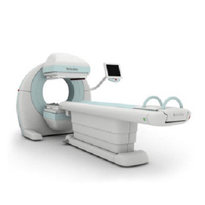
دستگاه SPECT

دستگاه SPECT

کاربرد SPECT

به کمک مقطع نگاری رایانه ای تک فوتونی (SPECT) ، می توان تصاویر سه بعدی از عملکرد یک بافت یا ارگان مانند قلب یا مغز، به دست آورد. روش کار به این صورت است که یک ماده با نیمع عمر کم ، به ترکیب شیمیایی خاصی تبدیل می شود که در بافت های خاصی از بدن و به نسبت فعالیت آن بخش،جذب خون می گردد.به عنوان مثال این ماده ی شیمیایی می تواند یک نوع قند خاص برای بررسی عملکرد بخشی از مغز باشد. این ماده وارد خون شخص شده و توسط سلول های مغزی که مصرف کننده اصلی این نوع قند هستند جذب می گردد و لذا بیشترین میزان تشعشع از این نواحی صورت خواهد گرفت.

اجزای دستگاه SPECT چیست؟

این دستگاه دارای یک تخت و یک فضای تونل مانند می باشد که داخل آن هد دستگاه قرار دارد. داخل هد اجزایی از جمله دوربین و آشکارساز قرار گرفته است که مراحل دریافت پرتوهای گاما و تبدیل آن ها به تصویر را انجام می‎دهند. هد این دستگاه  به صورت خودکار در زوایای مختلف به دور بیمار گردش مى کند و تصاویر متعددی از بدن بیمار را تهیه می‌کند. در حقیقت، دستگاه اسپکت به جمع آورى اطلاعات (پرتوهای ساطع شده از بدن بیمار) درمجموعه اى از جهت هاى مختلف می‌پردازد.


SPECT  چه خطراتی را در پی دارد؟

به طور كلی می توان گفت که انجام اسكن با اسپکت هیچ نوع خطری برای بدن ندارد؛ زیرا مقادیر بسیار کمی از مواد رادیو اکتیو در این روش استفاده می شوند. این مقدار دز تزریق شده با توجه به میزان دزی که سازمان بین المللی حفاظت در برابر اشعه برای یک انسان بالغ در مدت زمان مشخص تعیین کرده است؛ به هیچ وجه مشکلی برای بافت های بدن ایجاد نمی‎کند. ضمن اینکه با رعایت استانداردهای تصویربرداری با اسپکت، تا به حال هم هیچ مورد آسیب دیده‏‎ای با این روش تصویربرداری گزارش نشده است.
در تصویر زیر می توانید تفاوت کیفیت تصویربرداری SPECT را با MRI ملاحظه بفرمایید.


آمادگی‌های لازم برای این اسکن چیست؟

بیمار باید یک تا سه ساعت قبل از انجام اسکن، ناشتا باشد و مصرف یک سری از داروها را 48 ساعت قبل از انجام اسکن با مشورت پزشک قطع کند. به طور کلی 24 ساعت قبل از انجام اسکن موادی که محتوی کافئین هستند مانند چای، قهوه، نوشابه و شکلات نباید مصرف شوند.
توجه : بیماران آسمی صبح روز اسکن نباید از هیچ نوع اسپری دارویی استفاده کنند.
توجه : در رابطه با بیماران دیابتی می توان گفت که باید صبح روز اسکن از تزریق انسولین و مصرف قرص گلی بنکلامید و متفورمین خودداری کنند.


این مطلب را هم میتونی بخونی  تست سرخجه در زنان باردار

SPECT (single photon emission computed tomography) scan

Overview

A single photon emission computed tomography (SPECT) scan is an imaging test that shows how blood flows to tissues and organs. It may be used to help diagnose seizures, stroke, stress fractures, infections, and tumors in the spine.

How does a SPECT scan work?

SPECT is a nuclear imaging scan that integrates computed tomography (CT) and a radioactive tracer. The tracer is what allows doctors to see how blood flows to tissues and organs.
Before the SPECT scan, a tracer is injected into your bloodstream. The tracer is radiolabeled, meaning it emits gamma rays that can be detected by the CT scanner. The computer collects the information emitted by the gamma rays and displays it on the CT cross-sections. These cross-sections can be added back together to form a 3D image of your brain.
The radioisotopes typically used in SPECT to label tracers are iodine-123, technetium-99m, xenon-133, thallium-201, and fluorine-18. These radioactive forms of natural elements will pass through your body and be detected by the scanner. Various drugs and other chemicals can be labeled with these isotopes.
The type of tracer used depends on what your doctor wants to measure. For example, if your doctor is looking at a tumor, he or she might use radiolabled glucose (FDG) and watch how it is metabolized by the tumor.
The test differs from a PET scan in that the tracer stays in your blood stream rather than being absorbed by surrounding tissues, thereby limiting the images to areas where blood flows. SPECT scans are cheaper and more readily available than higher resolution PET scans.

What does a SPECT scan show?

A SPECT scan is primarily used to view how blood flows through arteries and veins in the brain. Tests have shown that it might be more sensitive to brain injury than either MRI or CT scanning because it can detect reduced blood flow to injured sites.
SPECT scanning is also useful for presurgical evaluation of medically uncontrolled seizures (Fig. 1). The test can be performed between seizures (interictal) or during a seizure (ictal) to determine blood flow to areas where the seizures originate.
Figure 1. A SPECT scan of a patient with uncontrolled complex partial seizures. The temporal lobe on the left side of the brain shows less blood flow than the right, confirming for the surgeon the nonfunctioning area of the brain causing seizures.
This type of scanning is also useful in diagnosing stress fractures in the spine (spondylolysis), blood deprived (ischemic) areas of brain following a stroke, and tumors.

Who performs the test?

A specially trained nuclear medicine technologist will perform the test in the Nuclear Medicine department of the hospital, or at an outpatient imaging center.

How should I prepare for the test?

Wear comfortable clothing and be prepared to stay for 1 to 2 hours.

What happens during the test?

First, you will receive an injection of a small amount of radioactive tracer. You’ll be asked to rest for about 10-20 minutes until the tracer reaches your brain. Next, you’ll lie comfortably on a scanner table while a special camera rotates around your head. Be sure to remain as still as possible so that the machine can take accurate pictures.
Once the scan is complete, you can leave. Be sure to drink plenty of fluids to flush the tracer from your body.

What are the risks?

The tracer is radioactive, which means your body is exposed to radiation. This exposure is limited, however, because the radioactive chemicals have short half-lives. They breakdown quickly and are removed from the body through the kidneys.
The long-term risk of radiation exposure is usually worth the benefits of diagnosing serious medical conditions. Your exposure risk could vary, however, depending on how many CT or other scans you have had. If you have concerns about your cumulative radiation exposure, talk to your doctor.
Women who are pregnant or nursing should not undergo a SPECT scan.
Some people may have an allergic reaction to the tracer or the contrast agent.

How do I get the test results?

The nuclear medicine doctor will promptly review your images and communicate directly with your referring doctor, who in turn will discuss the results with you.

Glossary

gamma rays: electromagnetic radiation emitted during radioactive decay and having an extremely short wavelength.
positron emission tomography (PET): a nuclear medicine test in which tissue function can be imaged. Damaged tissues have reduced metabolic activity; therefore, gamma radiation from these areas is reduced or absent.
photon: a particle that travels at the speed of light.
positron: an electrically charged particle that has the opposite charge as an electron. It reacts with an electron to produce gamma rays.
radiolabel: the technique of attaching, or “tagging”, a radioactive molecule to another molecule (such as a protein) so that it can be identified in the body. The radiolabeled substance emits positrons that can be picked up by a special scanner.
tomography: the technique of using rotating X-rays to capture an image at a particular depth in the body, bringing those structures into sharp focus while blurring structures at other depths.
tracer: a substance, usually radioactively labeled, which is injected into your body and can be followed to gain information about metabolic processes.
برای اطلاعات بیشتر در مورد دستگاه SPECT بر روی لینک زیر کلیک نمائید.

دیدگاهتان را بنویسید

نشانی ایمیل شما منتشر نخواهد شد. بخش‌های موردنیاز علامت‌گذاری شده‌اند *